Diagnostics for your eye health

Anterior and posterior eye
with state-of-the-art technology in view

The Sanoculus ophthalmologists in Berlin attach great importance to comprehensive and precise diagnostics in order to provide you as a patient with the best possible treatment and to preserve your vision in the long term.

We distinguish between special consultations such as contact lens fitting and myopia in children (myopia) and diagnostics of the anterior and posterior segments of the eye.
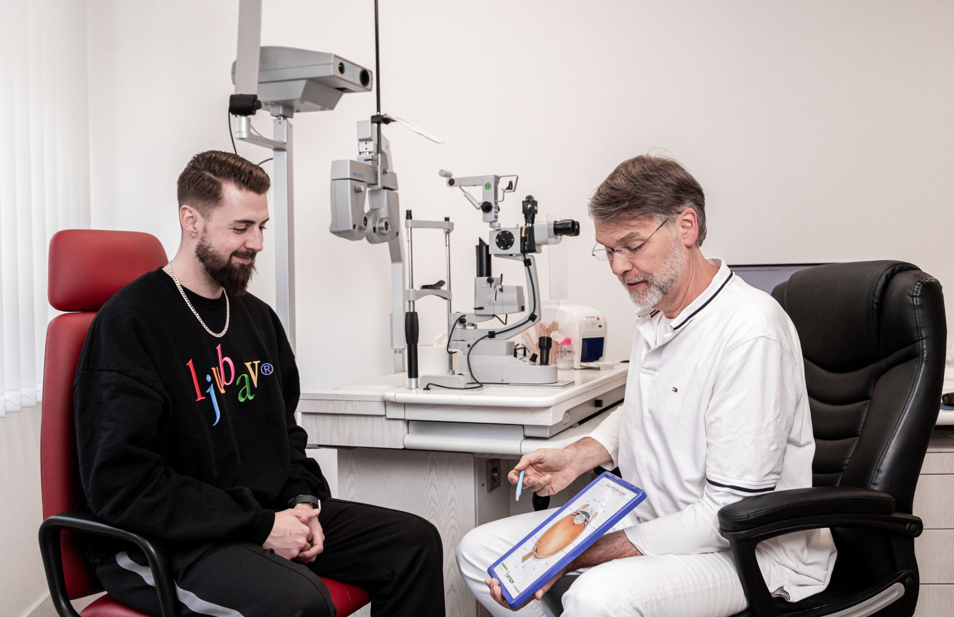
Augenarzt erklärt Patient das Auge anhand eines Schaubilds

Anterior eye

At the Sanoculus ophthalmologists in Berlin, comprehensive diagnostic examinations of the front eye are carried out. These include, among other things, the slit lamp examination, the examination of intraocular pressure and the assessment of the cornea and conjunctiva. With the help of state-of-the-art technologies such as optical coherence tomography (OCT), possible diseases such as conjunctivitis, keratitis or glaucoma can be detected at an early stage and treated effectively .

Posterior Eye

The diagnosis of the posterior segment of the eye also plays an important role for Sanoculus ophthalmologists. With the help of special examinations such as fundoscopy and OCT, diseases such as macular degeneration, diabetic retinopathy or retinal detachment can be detected at an early stage. Timely diagnosis and treatment can prevent or at least greatly slow down impending vision loss.
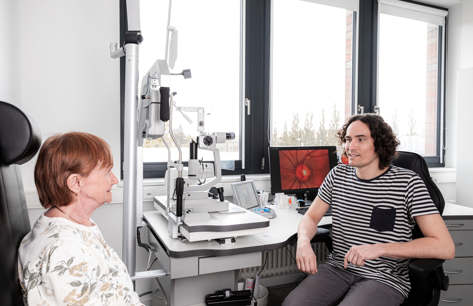
Augenarzt berät Patientin bei Sanoculus
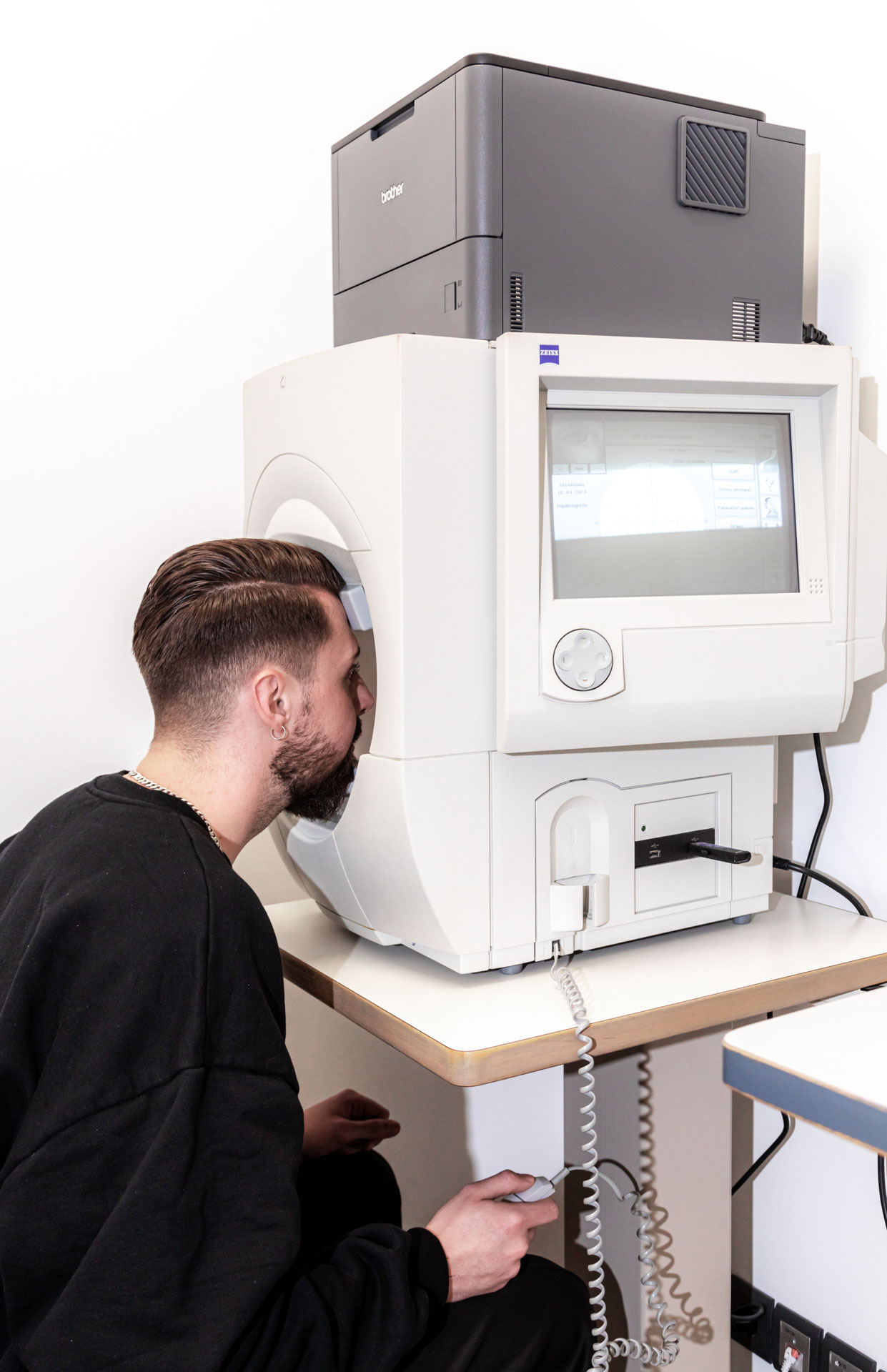
Patient schaut in

Visual field measurement / perimetry

Visual field measurement, also known as perimetry, is an important examination method to determine how well the optic nerve is functioning. Especially with high intraocular pressure, parts of the optic nerve can be damaged. Visual field measurement can detect losses in the peripheral visual area.

Especially in the case of glaucoma, it is important to have a visual field measurement carried out regularly. This is because vision loss occurs insidiously and is often not noticed by patients. Visual field measurement can detect vision loss at an early stage and localize it precisely. It is therefore advisable to include the visual field measurement in the routine examinations by the ophthalmologist. It offers the opportunity to detect any visual impairments at an early stage and to initiate appropriate measures. So have a visual field measurement done regularly to make sure that your optic nerve is intact and that you can continue to see well. Your ophthalmologist will be happy to provide you with all further information and support you in maintaining your eye health.

  1. Examples: -A patient notices that he suddenly has problems recognizing objects to the side while driving.
    A visual field measurement can be used to detect vision loss in this area.
  2. A patient notices that she suddenly can no longer recognize words at the edge of the page when reading.
    A visual field measurement shows that there has indeed been a loss of vision in the peripheral vision area.
  3. An elderly gentleman reports that he has difficulty perceiving fast-moving objects in his lateral field of vision when watching television.
    A visual field measurement can be used to identify visual loss in the field of vision.
  4. A woman notices that she is suddenly unsteady at night when moving around in dark rooms.
    A visual field measurement can detect any loss of vision in the dark.
  5. A patient complains that he suddenly can no longer see objects to the side of him during sport.
    The visual field measurement shows a restriction of the visual field in this area, which indicates a loss of vision.

OCT

Optical coherence tomography, or OCT for short, captures detailed cross-sectional images of the retina to assess the different layers of the fundus of the eye.
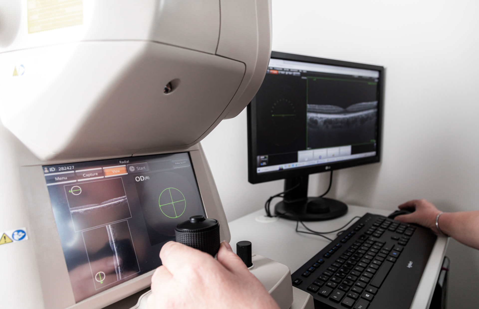
This examination is of great importance, especially in the case of age-related macular degeneration (AMD), in order to diagnose it early and monitor the course of the disease. In addition, OCT is also extremely valuable for other retinal diseases, such as diabetic retinopathy, retinal bleeding or fluid retention. In the case of glaucoma, OCT can be used to check whether there is already a change in the retina at the junction with the optic nerve, which can provide important information about the progression of the disease. It is therefore advisable to have an OCT examination carried out regularly if you have such eye problems or as a preventive measure. Your ophthalmologist will help you keep an eye on your eye health and take early action if necessary. Examples:

  1. Age-related macular degeneration (AMD): A patient suddenly notices blurred letters or a dark spot in the center of their field of vision when reading.
    These changes can be signs of AMD.
  2. Diabetic retinopathy: People with diabetes may notice sudden visual disturbances, such as fluctuating visual acuity or sudden flashes in the field of vision.
    These symptoms may indicate diabetic retinopathy.
  3. Retinal hemorrhages: A patient experiences sudden darkness or shadows in the field of vision that do not move.
    This could indicate a hemorrhage in the retina that requires urgent medical attention.
  4. Fluid retention: People might suddenly notice distortions or ripples in their field of vision, especially when looking at straight lines.
    This could indicate fluid retention in the retina, which can be diagnosed by an OCT scan.
Bildschirm mit OCT Werten
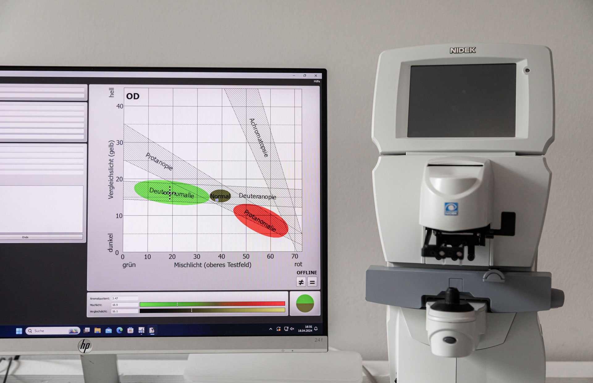
Messmonitor eines Anomaloskops

Anomaloscope

The Anomaloscope is a modern device specifically designed to detect color blindness , providing accurate and reliable diagnosis.

Additional information: Every tenth man has a red-green weakness and only every 100th woman.

With its innovative technology, color vision defects can be precisely determined and differentiated in order to offer those affected a better insight into their color perception.
For people who suffer from color blindness or have a suspicion, the anomaloscope is an indispensable solution to understand their vision and take appropriate action. Precise tests can be used to determine the type and severity of color vision deficiency in order to find individual treatment or correction options.

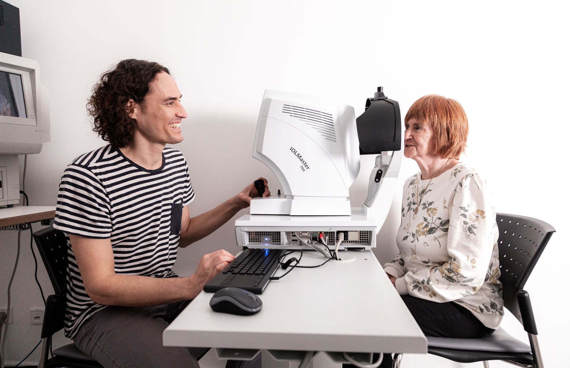
IOL Master

It is a measuring device that measures the entire eye very precisely. The following parts of the eye can be detected:

  • Cornea
  • Eyeball
  • Front chamber
  • Thickness of the crystalline lens

A measurement with the IOL master is carried out before cataract surgery . The exact measurement results can be used to determine the optimal strength of the artificial lens. The measurement is comfortable and contactless and does not require anesthesia of the eye. The patient looks into a device with his eye open and a beam of light quickly determines all the details of the eye. The IOL-Master sets new standards in measurement accuracy, in the past you had to perform an ultrasound of the eye, this was more unpleasant for the patient and the knife teeth were many times less accurate.

Augenarzt und Patientin sitzen sich beim IOL Master gegenüber
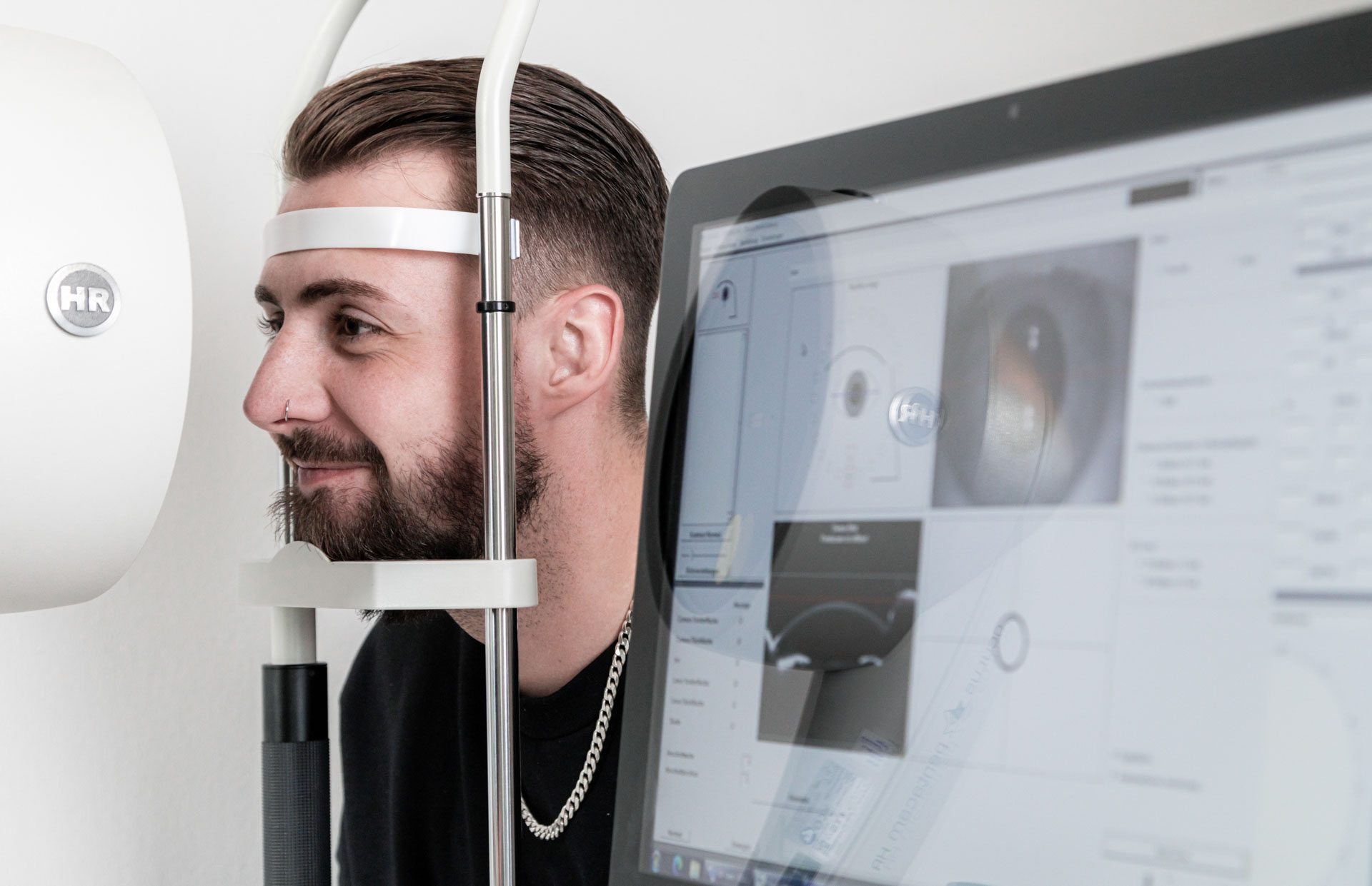
Patient schaut in Augenvermessungsgerät im Hintergrund. im Vordergrund werden die Ergebnisse auf einem Monitor angezeigt.

Pentacam

The image of a pentacam allows the visualization of the patient’s anterior segment of the eye. A Pentacam image is taken in preparation for cataract surgery .

In addition, the cornea can be precisely absorbed in different corneal diseases and serves as the basis for individual contact lens fitting. If keratoconus is present (irregular, cone-shaped deformation of the cornea), a Pentacam image is important to record the progression. Pentacam imaging is also advantageous in the case of glaucoma , as the anterior chamber angle can be determined and thus the form of glaucoma (e.g. narrow-angle glaucoma) can be defined. Admission with the Pantacam is painless, contactless and very pleasant for the patient. He looks into a measuring device and fixes on a red dot. With a laser, the Pantacam now takes your picture. No anesthesia or dilation of the pupil is necessary.

Endothelial cell microscope

The endothelium is the innermost layer, the five-layer cornea of the eye. In order to see clearly, it is important to have many intact endothelial cells.
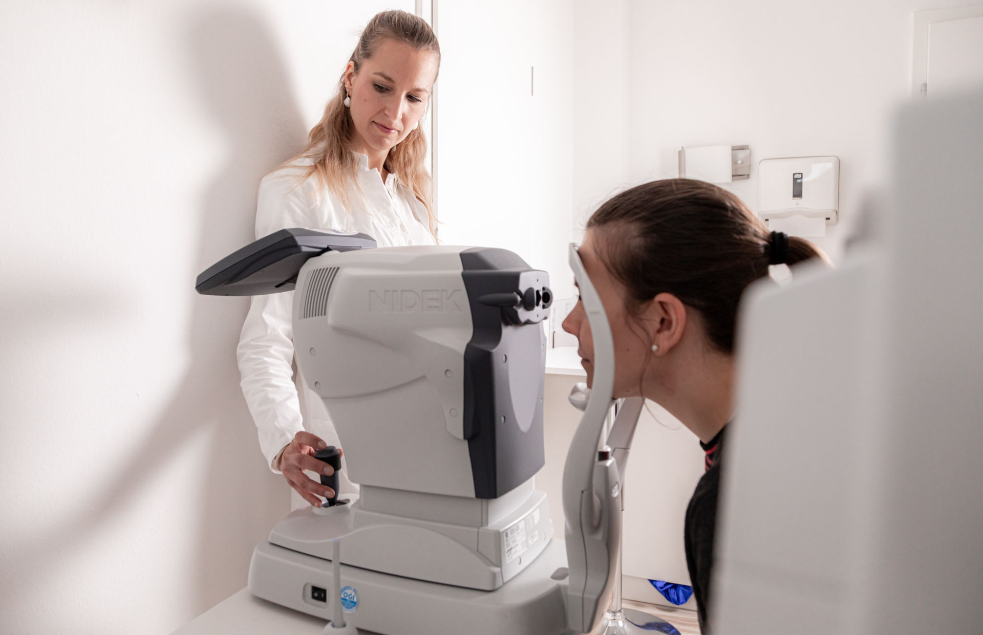
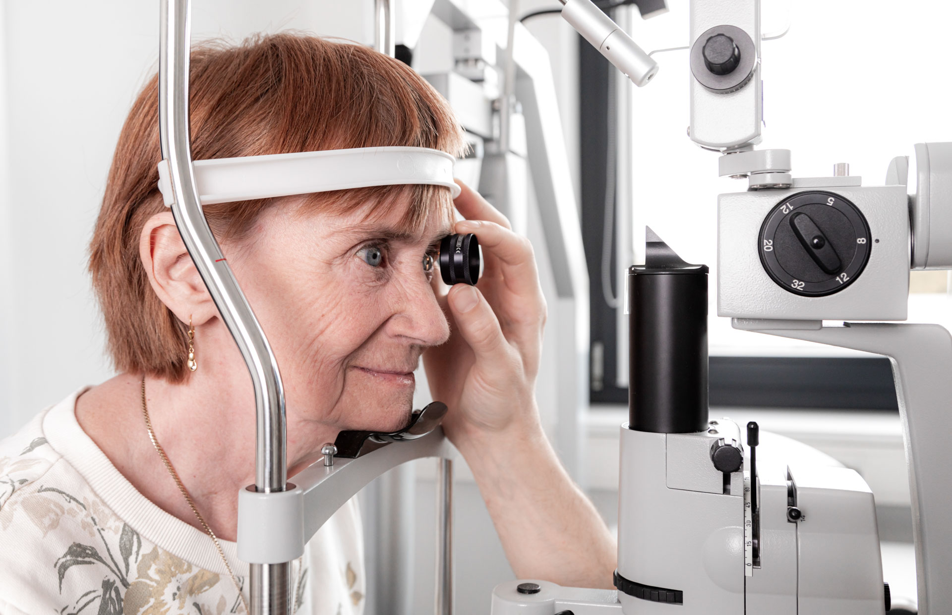
If there are too few endothelial cells, the cornea becomes cloudy and vision is impaired. The endothelial cell microscope is used to photograph the structure and number of endothelial cells. The following factors change the endothelial cells:

  • Surgery on the eye
  • Wearing contact lenses for too long
  • Medication
  • Chemicals
  • Corneal diseases
Augenärztin bei der Augenvermessung einer Patientin
Patientin schaut in ein Augenmessgerät

SLT Laser

SLT laser treatment for glaucoma is a method that works specifically and gently on the pigment cells of the eye in order to lower intraocular pressure.

The surrounding tissue remains intact, which makes the treatment minimally invasive and painless. The laser is used for less than 5 minutes to allow the aqueous humor to drain better and thus reduce the pressure in the eye.
With selective laser trabeculoplasty (SLT), the SLT laser offers an alternative to treatment with eye drops. By stimulating the outflow of aqueous humor, the SLT laser effectively lowers intraocular pressure. This low-risk treatment can simply be carried out on an outpatient basis.
SLT laser therapy often leads to such good results that no further eye drops are necessary. If necessary, the treatment can be repeated if the result is not satisfactory or if the intraocular pressure rises again over time. Thanks to this gentle and effective method, patients with glaucoma can achieve better eye health.

NCT (Non-Contact Tonometry)

Non-contact tonometry (NCT) is a pleasant and non-contact method for measuring intraocular pressure.
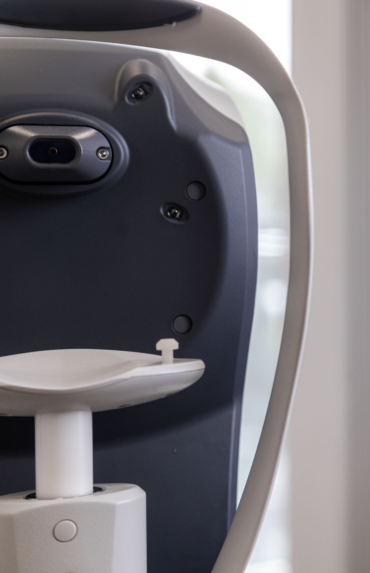
A slight barometric pressure measurement causes minimal deformation of the cornea, which is used by a measuring device to determine intraocular pressure. This is then combined with corneal thickness to determine the exact and true intraocular pressure. The determination of corneal thickness is also done with a special device.
Especially in the case of glaucoma or a familial risk as well as high blood pressure, it makes sense to have the intraocular pressure checked regularly. In our practice, we offer the possibility of measuring intraocular pressure using non-contact tonometry. A short puff of air is blown into the eye, and the reaction of the cornea is used to reliably determine the intraocular pressure.

Nahansicht eines NCT
Augenärztin bei der Vermessung der Augen eines Patienten

YAG Lasers

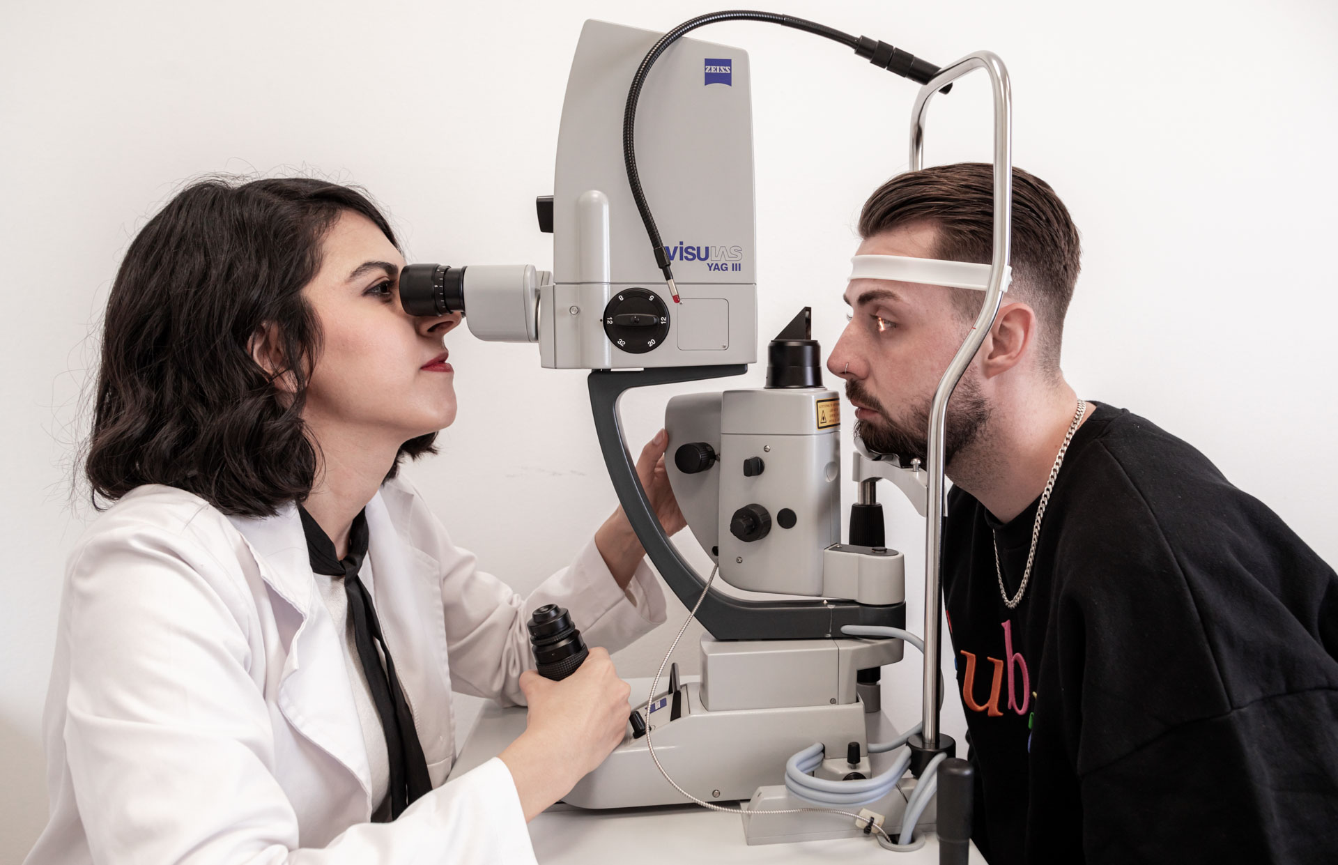
The YAG laser is used when a patient suffers from cataracts, which often occur as a result of cataracts.

After cataract surgery, the lens capsule left in the eye can become cloudy, leading to cataracts. This manifests itself in symptoms such as a decrease in visual acuity and a foggy, gray visual impression. The treatment of the post-cataract is carried out by the YAG laser without the need for further surgery. Local anesthesia of the eye is sufficient for this. By removing the membrane above the lens capsule with the YAG laser, clear vision is restored. The YAG laser (neodymium-doped yttrium-aluminum-garnet laser) is often used in the anterior eye area. In certain forms of glaucoma, such as narrow-angle glaucoma, the YAG laser can be used to create
a small opening in the iris.Summary: The YAG laser is successfully used to treat cataracts, which often occur after cataract surgery. The precise application of the YAG laser allows the cloudy membrane above the lens capsule to be removed, resulting in clear vision. In addition, the YAG laser can also be used to treat special forms of glaucoma, such as narrow-angle glaucoma.

FAG Laser

Fluorescence angiography (FAG) enables a detailed assessment of the fundus of the eye down to the finest vessels of the retina.
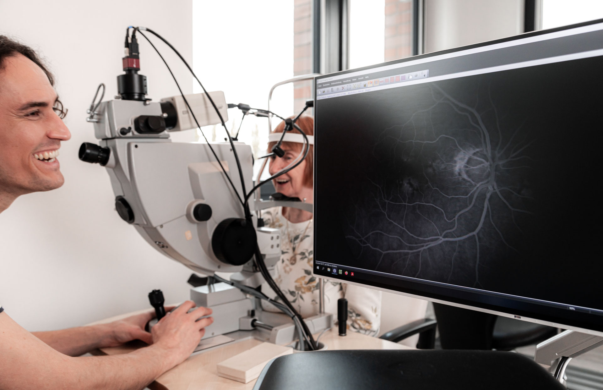
In this examination, a dye is usually injected that spreads into the smallest vessels of the retina. This allows eye diseases to be detected at an early stage and their progression to be observed. The FAG is particularly useful for the following clinical pictures, possibly also regularly:

  • Diabetic retinopathy (particularly important for diabetics)
  • Age-related macular degeneration (AMD)
  • Thrombosis (also possible in the retina)
  • Vascular occlusions and neoplasms
  • Tumors and inflammations

This contactless examination is carried out using a special camera that takes pictures of the fundus of the eye as soon as the dye reaches the retinal vessels. Sanoculus am Roseneck also offers FAG examinations, in which no dye has to be injected. This method, known as OCT-A, is especially beneficial for patients with chronic kidney disease.

Augenarzt und Patientin bwim FAG-Laser, Monitor mit Ergebnissen im Vordergrund

Stop myopia (myopia in children)

Myopia, also known as myopia, is a common vision problem in children. People with myopia have difficulty seeing distant objects clearly, while close objects remain clearly visible. In recent years, the incidence of myopia in children has increased.

The increase is partly due to changes in lifestyle factors such as increased screen use and less time outdoors. Early diagnosis and treatment of myopia is essential to slow the progression of myopia and reduce the risk of later complications such as retinal detachment or glaucoma. In special consultation hours at Sanoculus Ophthalmologists Berlin, myopia in children is specifically examined and individual therapeutic approaches are developed in order to respond to the needs of the young patients in the best possible way.

Have contact lenses fitted by an ophthalmologist

The use of contact lenses offers an excellent alternative to glasses, especially for patients who suffer from myopia or other refractive errors. In order to ensure optimal fitting and handling, the Sanoculus Ophthalmologists Roseneck offer special consultation hours for contact lenses .
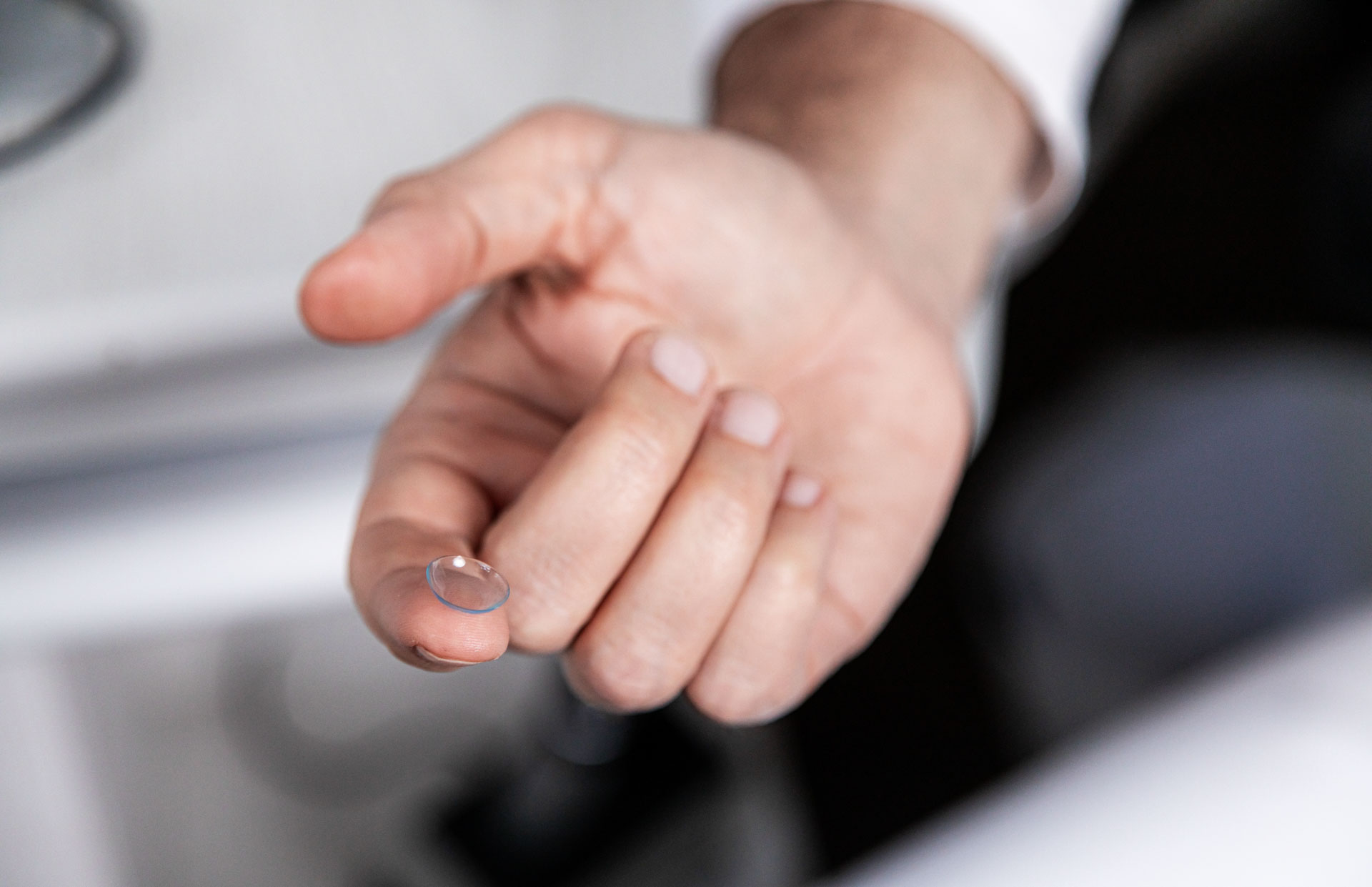
During these appointments, experienced ophthalmologists perform comprehensive consultations and adjustments to ensure that the contact lenses are perfectly matched to the individual visual needs and eye features of the patients. In addition, detailed instructions on the care and use of the contact lenses are given to maximize their comfort and safety. These specialized consultations ensure that each patient receives personalized care and can therefore experience the best vision and comfort.

Kontaktlinse auf einem Zeigefinger

OP-Center Sanoculus Elsterplatz Kissinger Str. 1 14199 Berlin

OP-Center Sanoculus Spandau Wilhelmstr.
23a 13593 Berlin

Sanoculus Roseneck ophthalmology practice Rheinbabenallee 12 14199 Berlin

Sanoculus ophthalmology practice Charlottenburg Paulsborner Str. 2 10709 Berlin

Ophthalmology Practice Sanoculus Kurfürstendamm Kurfürstendamm 115B 10711 Berlin

Sanoculus Haselhorst ophthalmology practice Burscheider Weg 11G 13599 Berlin

Sanoculus Steglitz ophthalmology practice Albrechtstr.
99 12167 Berlin

Sanoculus Rudow ophthalmology practice Köpenicker Str. 184 12355 Berlin